3d Printing in dentistry – Owandy Radiology
13 January 2025Guided implant surgery
15 January 2025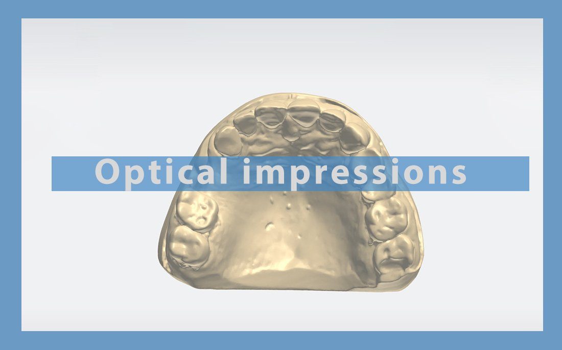
Optical impressions in odontology
Introduction to optical impressions
Optical impressions first appeared in the 1980s. They have now replaced conventional impressions, which were affected by various clear and recognised limitations, including stretching of impression materials, tearing, air bubbles that compromised the quality of these impressions and timeframes, as well as all processing phases required from the moulding stage, which considerably increased the number of adjustments in the mouth, and the risk of the final prosthesis failing.
Since their invention, intra-oral scanners and cameras have undergone numerous developments and are now an integral part of dental surgeries, their use indicated in all practices as well as in various odontology fields.
Definition
Optical impressions are defined as being a projected light signal onto dental surfaces. The reflected signal will be recaptured by the camera, allowing it to create the 3D model.
Optical impressions are the first link in the dental digital chain
Using optical impressions is an initial stage in data acquisition, followed by a phase in which this data is processed.
This can be done either by:
Scanning the model in the laboratory using an extraoral scanner or tabletop scanner (with or without contact), which scans a plaster model from an impression or straight from a traditional physical impression.
An intra-oral camera placed directly in the mouth.
Various types of optical impressions and acquisition techniques
1-Extraoral acquisition
This is an automated indirect technique used to control the positioning, distance, speed and accessibility of undercuts.
2-Intraoral acquisition
This is a direct impression technique. It involves manual operation, so is operator-dependent, which makes it impossible to accurately control the distance, speed and accessibility of undercuts.
3D imaging using the intraoral scanner can be done either by recording successive shots, the main disadvantage of which is the acquisition speed, or by using dynamic mode (film type). In this case, acquisition will be much quicker (around 5 mins to scan both arches and occlusion).
Digital processing of the optical impression:
The file produced by dental surface acquisitions constitutes the entry point for the dental CAD/CAM chain.This comprises a representation of the mould or the patient’s arch in the form of a point cloud. This point cloud is not actually useable, and requires processing by dedicated software to optimise it by resetting, repositioning and merging the overlaid points, cleaning and filtering of outliers, and standardising densities, so as to reduce the file size and accelerate the processing of virtual data.
Files obtained when exiting the acquisition link
These files can be in a proprietary format, which can only be read by design software of the same brand or a universal one. But the trend is currently towards opening up systems; universal files can be read by all design software.
Universal files are STL (Standard Tesselation Language or Standard Triangle Language) and PLY (Polygonal File Format).
The STL universal file defines a triangular mesh that will create a 3D object in black and white and which does not include colour or texture.
The PLY file consists of a collection of polygons that can transfer information such as transparency and colour. But not all software is yet able to support this file format.
Characteristics of intraoral cameras
There are several parameters to consider when choosing an intraoral camera. The technology selected determines access to certain settings (e.g. no powder, colour, continuous recording). The main characteristics of intra-oral cameras are listed below.
Ergonomics of the handpiece
Ergonomics include the size and shape of the camera, its weight, its handling, the necessary height at the tip, and the size of the head.
It also includes how well the camera balances, which is key in ensuring optimal stability and handling during the acquisition process.
A wide head means a larger acquisition field, but this can make it harder to access certain areas, especially those at the back.
The focal length is defined as the distance between the camera tip and the surfaces to record. Some brands require contact with the teeth, while others recommend a distance of over 10mm, which makes optical impression tricky when it comes to the posterior aspect, especially for disto-vestibular areas.
The most common and familiar way to operate it is as a “pen”, but there are also “gun” type cameras available.
Wireless camera versions have recently been developed that obviate the need for a cable, with all its constraints, and deliver greater freedom of movement.
Powder
This is used to standardise the light reflected onto the surfaces being recorded (enamel, filling and crown materials, etc.).
Some systems require powdering before the dental impression, in order to facilitate the recording of dental surfaces. Others simply recommend it.
Powder-free intraoral cameras can occasionally require a light coating for particularly shiny areas (metal restorations).
The trend, however, is for powder-free acquisitions.
Capture mode (image acquisition)
This can be an image or video mode, the only difference being the speed of acquisition, which is quicker in continuous mode as a film.
This acquisition method enables greater freedom of movement, but a manufacturer-prescribed scanning pathway must be adhered to.
Colour
The majority of systems can now be used to obtain master model in colour that are extremely realistic. This gives the 3D file a texture akin to dental tissue, and makes it easier to read various elements, including complex juxta-gingival preparation limits.
It is, however, possible to switch to a monochrome display at any time, as the monochrome model is the actual working model equivalent to the plaster model and which will be used to judge the quality of our impression. Both display modes are complementary.
Colouring
Some intraoral scanners can also reproduce tooth colour, thereby helping the practitioner to choose the right shade.
File format
Previously, we would talk about restricted, controlled access or open access files. Nowadays, systems are increasingly geared to exporting in open STL format, making it possible to work on impressions with any type of CAD software.
Software functionalities
All software relating to optical impression cameras make it possible to visualise the prosthetic space available and occlusal contacts.
Cost
It is difficult to evaluate the depreciation of an optical camera. It includes the purchase cost of the camera, as well as the computer unit, operating licences, software updates and maintenance fees.
Chairside
Some systems offer a complete CAD/CAM chain (acquisition, CAD, CAM) enabling the practitioner to move straight to CAD/CAM if desired.
Modularity / scalability
Buying an optical impression camera can also constitute the first stage of a switch to CAD/CAM, as all systems can now be used to export files in .STL format The practitioner can acquire the same brand of equipment or that from another manufacturer.
Optical impression precision and the concept of accuracy
The first question that comes to mind when we talk about optical impressions is: what do they contribute in terms of quality and accuracy compared with conventional techniques, and what is the most reliable technique in the various clinical situations?
Accuracy is the term used to describe the quality or precision of an impression.
It takes two criteria into account:
Correctness: the deviation rate measured between the actual dimensions of the object and the impression measurement.
Precision: the differences measured between results obtained using repeated measurements of the same object and the same impression system.
Partial arches
A number of factors can affect the quality of a clinical impression. Evaluating the quality of a digital impression involves analysing the marginal adaptation of prostheses made from two types of impressions (digital and conventional) using the same CAD/CAM manufacturing chains.
Generally speaking, the acceptable marginal adaptation of the prosthesis is below 120μm. In vitro studies show that the marginal adaptation values of ceramic crowns obtained using optical impressions are equivalent to, if not better than, those obtained using traditional impressions.
This has been confirmed in vivo for unitary or minor restorations with an average marginal adaptation value equal to 90μm.
This is also the case for partial inlay/onlay type restorations whose marginal adaptation values obtained using a digital impression are satisfactory.
Full arches
According to evidence cited in publications, the current recommendation is a conventional impression with elastomers, as this remains considerably superior to digital impressions for the purpose of full arches. It is also noted that accuracy is greater in the anterior sector than the posterior sector, with deviations in the horizontal plane and precision errors of up to 170μm.
It is the digital reconstitution phase used in assembling the images obtained to recreate the full arch that is singled out by several authors.
It would therefore seem that an accumulation of errors occurring in the image recording and digital processing stage is at the root of these significant distortions.
According to some authors, this means we could think about obtaining more reliable full arch impressions in the future if we can improve software rather than cameras.
Furthermore, according to others, it would seem that the number of preparations is key in altering the quality of digital impressions. The greater the number, the more significant the differences between the optical impression systems.
Generally speaking, digital impressions are considered to deliver a degree of accuracy that is sufficient, if not higher, than that delivered by traditional techniques, for unitary and minor restorations (up to 3 elements). In any case, conventional elastomer impressions remain the gold standard for recording full arches, particularly where there are large numbers of preparations.
Overdenture implants
It is difficult to come to a conclusion regarding the quality of digital impressions for implant-borne fixed prostheses, due to a lack of research on the subject. But some authors emphasise that there are several criteria to observe in order to achieve an impression of equivalent or higher quality to that of a standard technique, and that an extremely rigorous protocol should be applied. The quality of the impression will therefore be operator-dependent and experience-dependent.
Acquisition operating protocol
Once the teeth have had all humidity removed (saliva, blood, etc.) and any necessary powdering has been done, thinly applied with no clumps, the acquisition can begin.
It is important to adhere to the technique, duration, speed and scanning trajectory recommended by the manufacturer. Movement should be fluid and quick, and should gradually move away from the mouth, coordinating movements with what can be seen on the screen.
Preparation or working model shots should be taken of the widest preparation axis in order to be of use and show certain areas legibly. For conventional peripheral preparations, these zones are:
—the entire cervical limit and transition line
—cervical preparation
—the line demarcating the prepared dental area
—the taste buds and contact points of adjacent teeth
If the preparation is “opto-legible” and these zones are easily distinguishable, then CAD and CAM will be optimised. Otherwise, there is a strong risk of encountering difficulties at the insertion stage, because of badly managed contact points or poor cervical adaptation, and even under or over-bite problems.
Acquisition is a three-step process:
—Preparation impressions (working model)
—Impression of antagonist arch (antagonist model)
—Impression of occlusal relationships by scanning the vestibular side of the relevant occlusion sector. The software therefore puts both arches in MIP, either automatically or manually.
Advantages
The optical impression can be used to reduce work time compared with a conventional impression, particularly for sector scanning, and can help reduce discomfort experienced by patients who feel that traditional impressions are too aggressive.
In certain complex cases requiring multiple impressions, or with patients at high risk of nausea, acquisitions can be conducted gradually while still retaining the quality of the impression.
Furthermore, optical impressions are more straightforward than conventional impressions because of their objectivity, in the sense that they leave no room for chance or individual interpretation.
In fact, they enable immediate feedback on the teeth preparations so they can be validated prior to submission as well as considerably improving the quality and legibility of impressions sent to the laboratory.
In addition, traditional chemical-manual impressions are long processes that require a certain level of technical expertise and experience.
Unlike plaster impressions, which are heavy, unwieldy, difficult to store, non-reproducible, fragile and non-preservable, digital impressions are inalterable and invariable (as they cannot be worn out or deformed over time); they are also duplicable, easy to store, and transferable (digital media, secure servers, etc.).
Hence the benefit of switching to digital impressions by intraoral acquisition. Furthermore, they boast an accuracy level equivalent to and possibly even higher than that of conventional materials, particularly for unitary and minor prosthetic rehabilitations.
Lastly, optical impressions are an excellent communication tool, whether for explaining planned prosthetic work to patients or for the prosthetist. These exchanges take place in real time.
Disadvantages
Although digital dental impressions offer numerous advantages, there are limits to their use, mainly concerning the patient, the optical system and the digital data processing software.
Saliva flows can negatively affect intraoral scanner systems, while sulcus bleeding can hide some of the preparation limits.
Recording preparations would also be complicated by poor camera accessibility, particularly in the posterior region, essentially due to limited mouth opening, a preparation limit that was too deep, and movement by the patient.
It is often said that the camera only records what it sees. Optical impressions are more straightforward than traditional impressions, but they are also more rigorous.
Preparation techniques should be adapted to suit digital impressions, and gingival eviction techniques are strongly recommended.
During the digital acquisition process, there are two main dispersion levels:
—Accuracy of raw point clouds (particularly if model scanning is from a physical impression) compared with the scanned object.
—Processing of point clouds compared with the raw cloud.
Powder application is necessary, for some systems and in some cases, but it can interfere with the accuracy of the acquisition if it is applied in thick layers. The optimal thickness lies somewhere between 40µm and 60µm. Some cameras, i.e. those without powdering, recommend the use of a powder spray to speed up the acquisition process.
In vitro studies comparing digital impressions with conventional impressions have shown that both techniques are equally valid in terms of accuracy. However, conventional impressions have the advantage of causing fewer dispersions, and full arches are now difficult to establish with an intraoral camera.
How to choose your intraoral camera
When choosing the right intraoral scanner for dental surgeons, it’s a good idea to discuss the issue with the prosthetist, in order to select the model that best fits the practitioner’s expectations but which is also compatible with the laboratory chain. It will then be necessary to conduct a trial and learning phase so as to become familiar with this new equipment.
The practitioner can find the multitude of technical data quite bewildering and sometimes difficult to decipher, with the various descriptions by all the different manufacturers, as well as the latest developments: wireless systems, colour recognition, compact models, etc.
Before starting, the main elements to consider are:
— Cost
—Performance in terms of accuracy
—Technical features: acquisition speed and data processing timeframes (algorithm), sensor size (no. of pixels and acquisition area), focal length (field depth), mould / impression / dental arch digitisation
—Equipment ergonomics: acquisition time, with or without powdering
Generally, cameras complying with current standards are equipped with video stream and colour acquisition, and are powder-free.
Conclusion
Optical impressions are a booming sector, and represent a viable alternative to conventional impression techniques.
They offer a reliable, quick and comfortable acquisition process that facilitates communication with prosthetics laboratories and patients, even though the quality of full arch scanning currently remains limited.
Having conquered the world of fixed prostheses, the industry is now focusing on removable prostheses, implantology and orthodontics so as to offer comprehensive solutions to dental surgeons who have converted to digital.